Arthroscopic Knee Procedures
Overview
Author: Dimitrios Giotikas MD, PhD
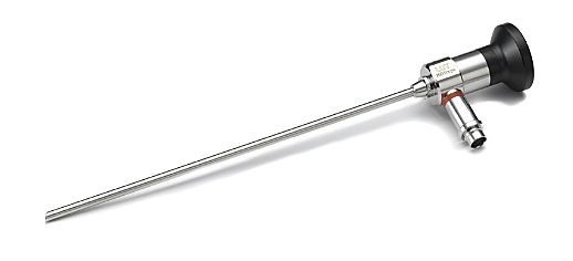
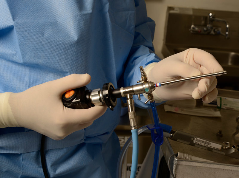
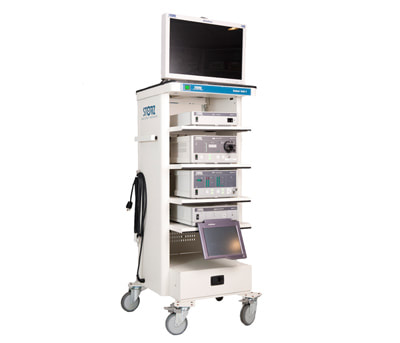
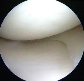
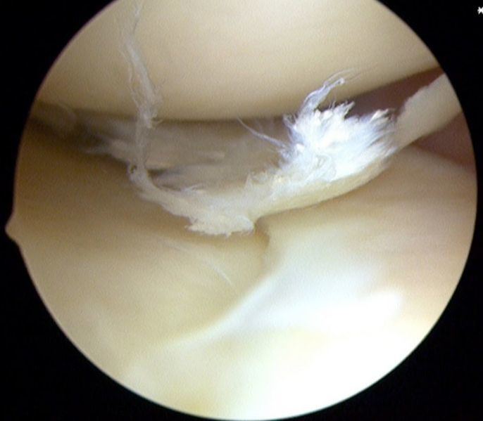
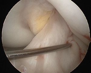
Knee arthroscopy is a surgical procedure where the surgeon inserts a thin (pen -like) arthroscope into the patient’s knee in order to diagnose and treat intra-articular problems of the joint. The arthroscope is connected to a camera and the images are transferred onto a screen. The surgeon sees real-time the intra-articular structures and lesions of the knee. Knee arthroscopy is performed through two small (stab) incisions, which are called portals, on the front of the knee. One of the portals is used for the insertion of the camera and the other for the insertion of instruments. Sometimes, some arthroscopic procedures may require one or two additional portals to allow access to specific areas of the knee joint. Because of its tiny access, in some countries arthroscopy is also referred to as “keyhole surgery”.
Arthroscopy has been a significant advancement in orthopaedics because it enabled doctors to better understand lesions and diseases in the joint and, equally importantly, to offer efficient treatments with minimal impact and morbidity. When knee arthroscopy first became available in the 70’s it was used primarily to look inside the knee joint and make a diagnosis. Today, knee arthroscopy is used in a wide range of different types of surgical procedures on the knee joint including confirming a diagnosis, removing loose bodies, removing,repairing or replacing a torn meniscus, reconstructing torn ligaments, repairing articular cartilage and fixing fractures of the joint surface.
Risks related with knee arthroscopy include:
- Infection
- Thromboembolism (Blood clots)
- Injury to nerves or vessels
- Persistent pain and knee stiffness
- Anaesthetic risks
- Accumulation of blood clots in the knee (haematoma)